Surgical Procedure

Figure 3: hypodermic needle placement

Figure 4: lag screw

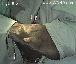
Figure 3: Under aseptic conditions, sterile hypodermic needles were placed in the proximal and distal intertarsal joints according to the landmarks established in Fig 2. This surgical procedure presents some challenges in orientation, and accurate preparatory steps are key to its success.

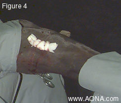
Figure 4: The lag screw is inserted through a stab incision on the craniomedial aspect of the joint between the needles shown in Figure 3. The short skin incision is protected by oversewing a "stent" bandage.

Figure 5: immediate postoperative radiograph

Figure 5: The immediate postoperative radiograph shows the 3.5mm AO ASIF cortex screw well-positioned in the central tarsal bone halfway between the proximal and the distal intertarsal joints. There appears to be good compression of the fracture line.

The small dark spot ahead of the tip of the screw indicates slight overdrilling of the thread hole. This is a good sign in a so-called "blind hole" since proceeding in this fashion eliminates the danger of the screw's "bottoming out". If this does occur, the implant may fail in torque, and the fracture, in any case, will not be compressed.

| Index Page | Case Presentation | Case Discussion | Case Follow-Up |

Case of the Quarter Index Page

AONA HOME PAGE