|
|
|
|
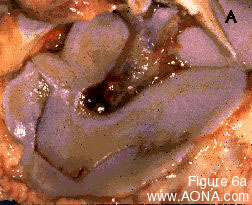
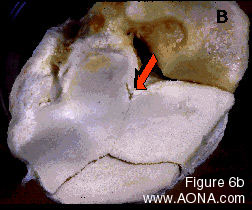
Figures 6 A & B: These two post mortem specimens illustrate two important problems associated with fractures of the cuboidal bones of the tarsus. In 6A, a fracture that appeared to be a simple slab radiographically is revealed to have a much more complex geometry (central tarsal bone viewed from its distal aspect). In 6B the relatively simple slab is seen to be complicated by a hairline crack in the bone (arrow), which is very close to the path a compression screw might take during repair. |
|
|
|
|
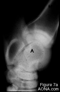
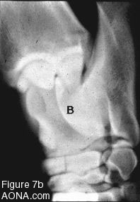
Figures 7 A & B: The fracture seen in 7A corresponds to the type of fracture seen in Fig 6A. It is impossible to fully evaluate the existing pathology. In 7B the presence of the additional "hairline" seen in 6B cannot be appreciated. CT scanning would be the only way to fully visualize these complexities. |