The history of right hindlimb lameness in this 14 year old Warmblood gelding used for dressage stretched over almost a year at the time of presentation. At times pain seemed severe, but would subside with rest and reduced intensity of exercise. There were never any obvious signs of local swelling, but the hock flexion test remained strongly positive, and diagnostic nerve blocks in the distal limb were ineffectual.
|
|
|
|
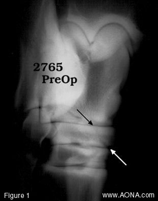
Figure 1: Although earlier radiographs were negative, this film taken in October 1997 shows an oblique fracture line (arrows) separating a slab-shaped fragment from the face of the central tarsal bone. |
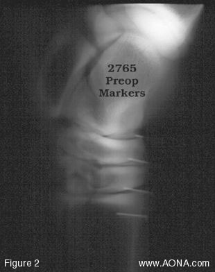
Figure 2: Our success in treating slab fractures of the tarsus by conservative means had been low, most animals developing chronic degenerative joint disease, and some suffering catastrophic collapse of the affected bone. We therefore recommended lag screw fixation. Preoperatively, radiopaque markers served as orientation. |