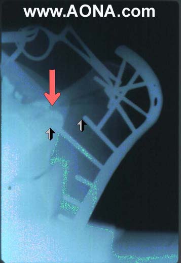
Figure 2: Initial Surgery - The immediate postoperative radiograph shows good contouring of the implant ( a 10 hole narrow dynamic compression plate), and compression of the transverse portion of the fracture. It also shows that the fracture had a vertical component (red arrow), and that the two screws that could have served to compress the fracture (split arrows) in fact do not reach it.