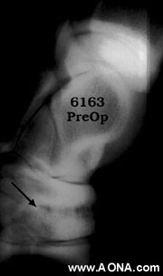
Figure 2
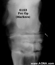
Figure 3
Due to the advanced stage of the disease, and the desire on the owner's part for a more permanent cure than is represented by antiinflammatory therapy, it was decided to perform surgical arthrodesis of the affected joints. Under general anesthesia and aseptic conditions, the patient was positioned inn left lateral recumbency, and the surgical site give the final prep. Following draping, a hypodermic needle was placed in the distal intertarsal joint (Figure 2). This serves to determine the placement of the proximal edge of the T-shaped compression plate (Figure 3).
Figure 3
Although the plate appears "elevated" from the bone in these exposures, it is in reality in tight opposition to the layer of proliferative bone that coats the surface of the cuboidal bones of the tarsus, and the proximal extent of the metatarsus. Experience has shown that it is unnecessary to disturb this periosteal proliferative tissue to bring about fusion. In fact, the cosmetic result is improved by avoiding curettage.