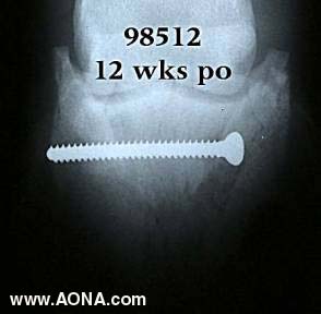
In this 12-week postoperative view, it may be seen that the fracture has begun to consolidate. The implant remains solidly in place, with no sign of osteolytic changes in the adjacent bone. The horse had been treated with the same corrective shoeing (bar-shoe with large side clips) as was in place prior to surgery.
At twenty weeks, the fracture has healed completely, and the screw may be removed. We have found this to be preferable to leaving the implant in place, since horses may suffer late-term (6 months - 1 year) complications. Those consist of slowly developing lytic changes around the screw head associated with recurring lameness. Occasionally there is associated seropurulent drainage.
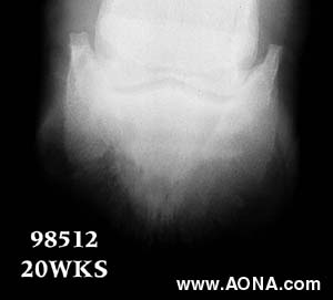
The technique employed for screw removal is similar to that described for its placement.
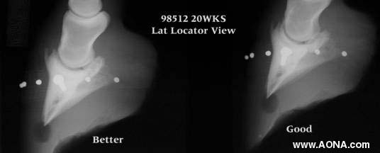
Radiopaque markers are affixed to the hoof wall prior to producing a lateromedial projection. It behooves one to be critical of positioning, lest the markers not truly reflect the internal anatomy. Scrutiny especially of the navicular bone in the film on the right shows some doubling of the margins of the bone, indicating it is not a true lateral.