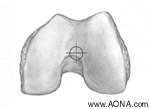
Axial view - insertion site
Click on image for enlarged view
Identify nail entry point
Make a medial parapatellar incision. Retract the patellar tendon laterally.
The entry point for the nail is in the intercondylar notch, just anterior and lateral to the femoral attachment of the posterior cruciate ligament. The location of the entry point in relation to the intercondylar notch varies with patient anatomy.

Click on image for enlarged view
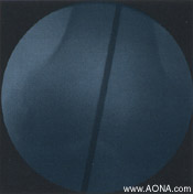
Thread the 13 mm/3.2 mm Trocar ]357.128] into the 13 mm Protection Sleeve [357.127] .Insert the assembly through the incision to the bone. Hold the Protection Sleeve firmly and pass the 3.2 mm Calibrated Guide Wire [292.69] through the trocar. Insert the guide wire using a power drill. The guide wire should be inserted in line with the anatomic axis of the femur which is 7°-9° lateral to a line perpendicular to the articular surface.


Click on images for enlarged views