DCS Surgical Technique, contd.
Click for enlarged view of image
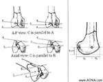
3. Using the DOS Drill Guide, insert the central guide pin
(C) parallel to the distal K-wire (A) in the A-P view, and
parallel to the anterior K-wire (B) in the axial view. Do not
insert the guide pin too far medially; consider the inclination
of the medial wall of the distal femur. In the sagittal plane,
the central guide pin enters the distal femur at a point
anterior to the midline between the condyles, and in line
with the shaft axis, approximately 2 cm from the knee joint.
Confirm placement of the central guide pin under image
intensification. If it is not parallel to the knee joint axis,
insert a new DHS/DCS Guide Pin.
Notes:
Because it is designed for use with the DHS/DCS instru-
ments and implants, the DHS/DCS Guide Pin, and not an
alternate pin, must be used.
This guide pin remains in place throughout the procedure.
If it is inadvertently withdrawn, reinsert it immediately.
(See "Reinserting the DHS/DCS Guide Pin")
Next Page
DHS / DCS Index